PET Scan
NUCLEAR MEDICINE SERVICES at AIC
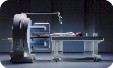
AIC has the valley's most sophisticated nuclear camera.

Q. Can you tell me more about the new NUCLEAR/SPECT/PET camera
at AIC?
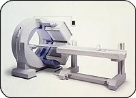
A. The Nuclear scanner at AIC (shown above) is the most
sophisticated in the Antelope Valley area. It is a top-of-the-line Siemens
state-of-the-art dual-head gamma camera that can perform full range
of Nuclear Medicine scans including SPECT scans as well as
PET scans.
 Q. What types of Nuclear studies are offered at AIC?
A. Full nuclear services are offered including bone
scans, HIDA scans, V/Q scans, thyroid scans, Indium white cell scans,
Gallium scans, brain SPECT, scintilymphangiography, cardiac scans
(thallium and Sestamibi SPECT, etc.).
Q. What is SPECT and what are some of the indications?
A. SPECT stands for Single Photon Emission
Computerized Tomography. SPECT is to planar nuclear scanning as CT
(CAT scan) is to planar x-rays. SPECT images are tomographic slices
through the region of interest. It is more sensitive than planar scanning
due to its finer thickness. Multiplanar imaging (coronal, sagittal, axial)
is available. SPECT is now the standard technique in cardiac imaging, but
has broad applications such as in bone scanning. Images on the right show
a coronal CT reformation of the spine demonstrating a sclerotic lesion in
T12 and the corresponding coronal SPECT image clearly showing increased
activity in T12 (arrows). The planar whole body
bone scan (not shown) was negative failing to show
the T12 lesion.
Q. What is a PET scan and what are some of the indications?
A. PET stands for Positron Emission Tomography.
PET imaging uses a glucose analogue agent called
F18-Fluoro-Deoxy-Glucose or FDG. In the body, PET is used in
oncology to detect malignancy since malignant cells demonstrate higher
glucose metabolism. Thus it can differentiate between malignant and benign
tumors and between recurrent tumor and scar tissue/radiation changes, etc.
In the brain, it can be used for malignancy as well as
Seizures/Epilepsy, Alzheimer and other Dementias, Schizophrenia,
Depression, Attention Deficit Disorder (ADD), etc. The images on the right
show a reformatted coronal CT image with the corresponding coronal PET
image of an unsuspected destructive metastasis to the left upper rib cage
(arrows).
Q. What agent is used in PET imaging?
A. PET imaging uses a glucose analogue called
F18-Fluoro-Deoxy-Glucose or FDG.
Q. What are some of the indications?
A. In the body, PET is used in oncology to detect malignancy
based on the fact that malignant cells demonstrate higher glucose
metabolism. Thus it can differentiate between malignant and benign tumors
and between recurrent tumor and scar tissue/radiation changes, radiation
necrosis, etc. In the brain, it can be used for malignancy as well as
Seizures, Alzheimer, Depression, Attention Deficit Disorder (ADD), etc.
Q. Does Medicare pay for PET?
A. Yes. Medicare currently pays for 5 indications related to
oncology applications:
- Solitary Pulmonary Nodule (SPN) ... may save the patient a biopsy
or surgery.
- Staging of lung cancer.
- Staging of Lymphoma.
- Staging of Melanoma and detection of recurrence.
- Recurrent colorectal cancer.
Private insurances pay for more indications including CNS
applications. All cases must be preauthorized, however.
AIC's nuclear/PET camera is unsurpassed in the
Antelope Valley area.
| ScanHealth |
Open MRI |
High-field MRI | MR Angiography |
Helical CT |
CT Angiography | Calcium Scoring
|
| 4D CT Reconstruction |
Dental Scan | 4D
Ultrasound | Nuclear Medicine | PET
Scan | DEXA Bone Density |
X-ray |

 Q. What types of Nuclear studies are offered at AIC?


 1.
The only community-based, private-practice, physician-operated
imaging facility in the Antelope Valley, just like any other private
practice medical office. Not belonging to any hospital or outside
imaging network. This means more personal and caring service.