|
|
||||
|
procedures |
home patients referring doctors 3-d image gallery locations contact us
|
|||
|
|
4D Ultrasound/Doppler 4D/3D ULTRASOUND
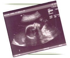
This amazing new technology is particularly useful in the OB patients for 4D visualization of the fetus (fetal face and other body parts, such as shown above). The mother will be given at least one 4D picture of the fetus at the end of the exam. Other applications include multiplanar imaging of the pelvic organs (including endovaginal 4D imaging). Other applications include multi-planar imaging (similar to MRI) of other body parts including abdomen, pelvis, small parts, neck, etc. We have a full-time sonographer on the premises and offer a full range of ultrasound services including: abdominal, pelvic, OB, head & neck, small parts, breast, and vascular Doppler (carotid Duplex/CVP's, venous/DVT's, renal Doppler, peripheral arterial/venous Doppler, etc.). A DVD with MUSIC is provided for the pregnant mom showing live 3D movie of her unborn baby.
ULTRASOUND
In medicine, ultrasonic waves are directed at a specific area of the body. As the waves travel through body tissues, they are reflected back at any point where there is a variation in tissue density, as in the area between two different organs of the body. The reflected waves are received by an electronic device that determines both the position of the tissue giving rise to the echoes and the intensity of the echoes. The resulting images can be displayed in static form, or, through the use of rapid multiple scans, they can in effect provide a moving picture of the inside of the body. It is a reliable, cost effective means of evaluating many internal organs including the liver, pancreas, spleen, kidneys, aorta, gallbladder, ovaries, uterus, prostate, testicles, and thyroid. Vascular ultrasound provides accurate assessment of arteries and veins in the extremities and neck. It is routinely used to evaluate fetal growth and complications of pregnancy. Ultrasound is particularly accurate for detecting gallstones, liver abnormalities, obstructed kidneys, vascular aneurysms, abnormalities of the uterus and ovaries, and tumors within the thyroid gland or testicles. Ultrasound is less accurate in very large patients. Ultrasound is very patient friendly -- there are no injections. Only a small instrument (transducer) will be in touch with the body. The only preparation for some exams is not to eat 4 hours prior, and for some pelvic exams, to have a full bladder. PRICING Silver Session 4D Sonogram Gender Determination Glossy color pictures 20 minute session $50 off Platinum session your second visit $100
Gold Session 4D Sonogram Gender Determination Glossy Color Pictures CD-ROM with Digital Color Pictures 30 minute session $50 off Platinum session your second visit $150
Platinum Session 4D Sonogram Gender Determination Glossy Color Pictures CD-ROM with Digital Color Pictures DVD of session set to music 45 minute session $200 Please be advised that individual results may vary depending upon maternal body habitus, fetal position, and gestational age. Recommended gestational age for 4D ultrasound is between 28 to 32 weeks. Discounted rates for multiple visits only applicable to same pregnancy. AIC is the most sophisticated and up-to-date medical
imaging center in the Antelope Valley and among the top facilities in
Southern California. We will continue to provide the best quality of service
possible for our physicians and their patients and constantly strive to
introduce the most advanced diagnostic medical technology to the Antelope
Valley community. We always welcome your comments for improving our
services. | ScanHealth |
Open MRI | High-field
MRI | MR Angiography |
Helical CT | CT
Angiography | Calcium Scoring
|
|
|||
|
copyright © 2004 ray h. hashemi, m.d., ph.d. |
||||


 Advanced Imaging Center is
proud to offer new revolutionary four-dimensional (4D) Ultrasound
machines (award-winning GE Voluson 730
Expert scanners) with on-line real-time 4D capabilities. It is the same
type featured on the Oprah television talk show and currently
being used for example at Cedars Sinai Medical Center.
  Diagnostic ultrasound is an established method of diagnostic medical imaging
using a high frequency sound wave and the principle of sonar.
Because ultrasonic waves cause no damage to human tissues, they are an
important tool utilized for both diagnosis and treatment of disease.
New generation ultrasound equipment provides images of very high resolution
and diagnostic accuracy.
  1.
The only community-based, private-practice, physician-operated imaging
facility in the Antelope Valley, just like any other private practice
medical office. Not belonging to any hospital or outside
imaging network. This means more personal and caring
service.