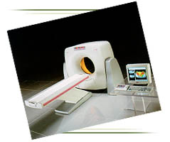
Three dimensional imaging, performed by our Toshiba
16-slice Helical CT which acquires data volumetrically, allows computer
processing to display various organs and disease processes three
dimensionally. This is now feasible because the volumetric scan data is
free of motion artifact and the low cost of computer power combined with
three dimensional software can create images previously unobtainable.
This process has many applications.
CT Angiography
The imaging of blood vessels in detail comparable to
catheter angiography at a fraction of the time and cost is a remarkable
advance. This technique will replace, to a great extent, invasive
angiography.
Musculoskeletal
Imaging of bones and joints three dimensionally
allows a different perspective for understanding pathology such as
complex fractures. It is very helpful for surgical planning.
Therapy Planning
Surgical and radiation treatment procedures are
complex three dimensional problems. Three dimensional rendering of CT
data is helpful for surgical planning and for the radiation oncologist.
For both, understanding the relationship of tumors to normal tissue and
blood vessels is critical. These imaging techniques are providing
information not previously obtainable.
Volume Rendering
Volume rendering is the most recent development. It
is a method by which the viewer is provided with a three dimensional
perspective of hollow organs. This technique known as virtual endoscopy
will be useful to image the trachea, bronchi, and the colon. Virtual
endoscopy of the colon is performed after a thorough colon cleansing and
distention of the colon with air. The helical CT scan data is then
processed to allow the viewer a three dimensional view of the inside of
the colon similar to that from an endoscope. This may someday play a
role in colon cancer screening.
"All Inclusive" Comprehensive Imaging
Three dimensional helical CT in many cases can now
provide all the information necessary for definitive treatment which
previously required two or more radiological studies. Its ability to
display both the pathology within an organ and blood supply to the organ
is unique and provides the most information for the least cost.

 1.
The only community-based, private-practice, physician-operated
imaging facility in the Antelope Valley, just like any other private
practice medical office. Not belonging to any hospital or outside
imaging network. This means more personal and caring service.